Cambridge Scientific
Cambridge Scientific Labs has New Biotech Space for Rent
Call 617-354-8900 for help! | Contact Us | About Us
Showing 1–12 of 25 results
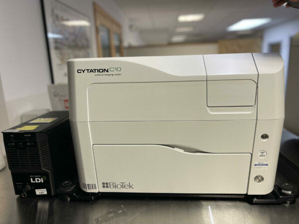
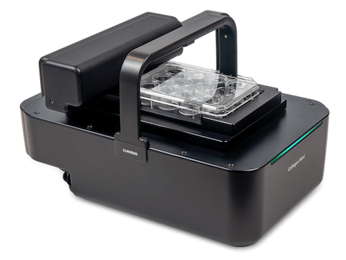
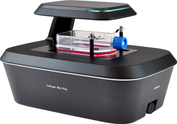
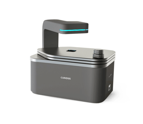
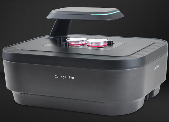
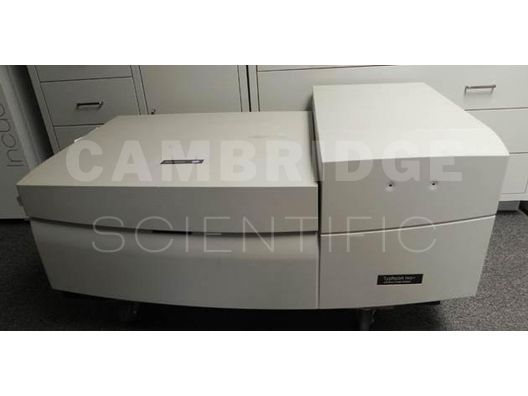
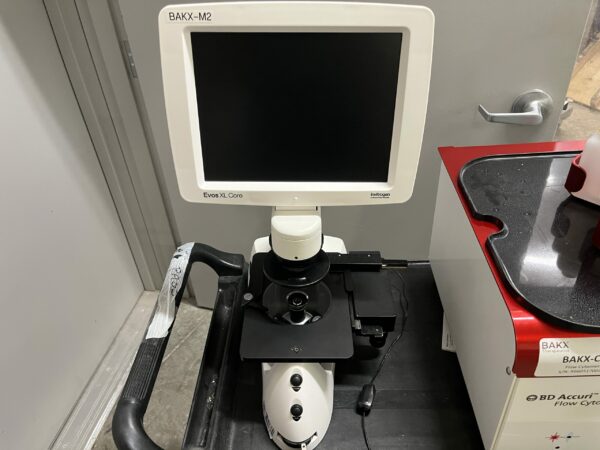
Product Type Select a product type -140 Chest Freezer -20 Freezer -20 Single Door Freezer -25 Freezer -30 Freezer -40 Freezer -80 chest freezer -80 Freezer -86 Freezer 3D Laser Scanning Microscope 3D Printer 4C Refrigeration Advanced Fluorescence Cell Analyzer Air Compressor Air Filtration System Alpha Blocks Analytical Balance Analytical Electronic Balance Analyzers Anti Vibration Marble Balance Table Antivibration Table Auto Lamination System Autoclaves Automated Cell Counter Automated Electrophoresis Automated Microplate Stacker Automated Microscope Automated Sample Storage Automatic Feeder Autostainer Axiophot Microscope Bead Bath Benchtop Autoclave Benchtop Centrifuge Benchtop Environmental Shaker Benchtop Lab Oven Benchtop Lyophilizers Benchtop Orbital Shaker Benchtop Pipette System Benchtop Platform Shaker Benchtop Refrigerated Centrifuge Benchtop Refrigerated/ Heated Centrifuge Benchtop Vacuum Concentrator Bioanalyzer Biological Microscope Biologics Analysis System Bioreactor Biosafety Cabinet Biosafety Cabinets BOD Incubator Camera Carbon and Sulfur Analyzer Casework Cassette Labeler CCD Microscope Monochrome Camera Cell Analyzer Cell Counter Cell Disruptor Cell Imaging System Cell Processing System Cell Sorter Cell Viability Analyzer Centrifugal Evaporator Centrifuge Centrifuges Chemistry Analyzer Chest Freezer Chiller Chromatography Chromatography Refrigerator Circulating Water Bath Clean Air Enclosure Clean Benches CO2 CO2 Incubator CO2 Water Jacketed Incubator Column Selector Compact Balance Compact Centrifuge Complete Culture Control Incubator Compound Fluorescent Microscope Compound Material Microscope Compound Microscope Concentrator Confocal Imaging Reader Confocal System Console Freeze Dryer Console Lyophilizer with Stoppering Console Stoppering Freeze Dryer Controlled-Rate Freezer Controller Convection Incubator Cooling Incubator Cover Slipper Cryo Storage Tank Cryogenic Cytocentrifuge Cytometer Dehumidifier Desiccator Dewar Diaphram Pump Digital Microscope Dissolution Apparatus Dissolved Oxygen Monitor DNA Analyzer DNA Sequencer DNA Synthesizer Double Door Refrigerator Droplet Digital PCR Drosophila Refrigerated Incubator Dry Bath Incubator Dry Block Heater Dry Blot System Dry Heat Sterilizer Dry Vacuum Pump Dual Convection Incubator Ductless Fume Hood Electronic Contact Thermometer Electronic Pipette Electronic Pipette Stand Electrophoresis Power Supply Electrophoresis System Electroporation System Endotoxing Testing System Environmental Chamber Evaporator Filtration System Flammable Cabinet Flammable Liquid Storage Cabinet Flammable Material Storage Refrigerator Flash Chromatography System Floor Incubator Shaker Floor Low Speed Centrifuge Floor Super Speed Centrifuge Floor Ultra Speed Centrifuge Flow Cytometer Fluidics Station Fluorescence Cell Analyzer Fluorometer Forced Air Incubator Forensic Microscope FPLC Autosampler FPLC Fraction Collector FPLC System Freeze Dryer Freeze Dryer w/ Stoppering FT-IR Spectrometer FT-IR Spectrophotometer FT-NIR Spectrophotometer Fume Hood Fumehood base Furnace Oven Fusion Glass kiln Gas Chromatography GC/MS Gel Documentation System Gel Imaging System Gene Synthesizer General Purpose Dual Chamber General Purpose Incubator General Purpose Water Bath Genome Sequencer Glass Door Lab Refrigerator Glassware Dryer Glassware Washer Glove Box Gravity Convection Incubator Gravity Convection incubators Gravity Oven Heat Block Heat Sealer Heated / General Purpose Waterbath Heated Circulator Heated Microplate Mixer Heated Water Bath Heated/ Circulating Waterbath Heated/ Refrigerated Circulator Heating Block High Capacity Centrifuge High Content Scanning System High End Research/Fluorescence Microscope High Intensity Illuminator High Speed Barcode Reader High Throughput Extraction System Homogenizer Hot Plate Hot Plate/Stirring Hot Plate HPLC Absorbance Detector HPLC Autosampler HPLC Autosampler Chiller / Thermostat HPLC Binary Pump HPLC Capillary Pump HPLC Column Compartment HPLC Degasser HPLC Detector HPLC Diode Array Detector HPLC Evaporative Light Scattering Detector HPLC Flex Cube HPLC Fluorescence Detector HPLC Fraction Collector HPLC Isocratic Pump HPLC Multi-Wavelength Detector HPLC Multicolumn Thermostat HPLC Multisampler HPLC Nano Pump HPLC Preparative Autosampler HPLC Preparative Pump HPLC Preparative System HPLC Pump HPLC Quaternary Pump HPLC Rack Changer HPLC Refractive Index Detector HPLC System HPLC UV/VIS Detector HPLC Variable Wavelength Detector HPLC Vialsampler HPLC Well Plate Autosampler Humidity Chamber Hybex Microsample Incubator Hybridization Oven Hybridization System Ice Flaker ICP-MS Imager Immersion Circulator Immunochemistry Analyzer Incubated Shaker Incubator Incubator Shaker Infared Imaging System Infrared Microsterilizer Integrated SpeedVac System INVERTED MICROSCOPE Inverted Phase Contrast Inverted Phase Contrast Microscope Inverted Phase Contrast/Tissue Culture Microscopes Inverted Research Phase Contrast Microscope Ion Chromatography Lab Bench Organizer Lab Freezer Lab Refrigerator Lab Stool Lab Table Laminar Flow Laminar Flow Hood Large Capacity Stirrer LC/MS System LC/MS Trap System LC/MS/MS Q Trap System LC/MS/MS System Light Source Liquid Dispenser Liquid Handler Liquid Handling System Liquid Nitrogen Dewar Liquid Nitrogen Storage Live Tissue Microtome LoadCell Controller Low Temperature Incubator Low-Pressure Chromatography System Magnetic Stirrer Maldi TOF MS System Mass Spectrometer Maxi Rotator Mixer Shaker Mechanical Convection Incubator Mechanical Convection Oven Mechanical Forced Air Incubator Mechanical Forced Air Oven Metabolic Analyzer Metabolite Analyzer Micro Balance Micro Bead Sterilizer MicroArray Scanner Microcentrifuge Microplate Absorbance Reader Microplate Bucket Microplate Carrier Microplate Centrifuge Microplate Fluorescence Readers Microplate Reader Microplate Shaker Microplate Spectrophotometer Microplate UV/VIS Reader Microplate Washer Microplate Washers Microscope Boomstand Microscope Camera Microscope Fiber Optic Light Source Microscope Fluorescence Light Source Microscope LED Fluorescence Light Source Microscope Metal Halide Fluorescence Light Source Microscope Monochrome Camera Microscope Table Microtome Microtome Cryostat Mill Grinder Mini Centrifuge Mini Stirrer Mini Vortexer Mixer Mixers, Stirrers & Shakers Moisture Analyzer Multi-Tube Vortexer Multilabel Counter Multilabel Microplate Reader Multimode Microplate Reader Multiplex Reader MultiTherm Shaker Nano Coating System Nano Particle Assembler Natural Convection Incubator Natural Convection Oven NIR Spectrometer Nitrogen Generator Nucleic Acid Automated Purification System Nucleic Acid Extractor Open Air Shaker Optics Table Orbital Shaker Osmometer Oven Overhead Stirrer Oxygen Monitor Paramount Filtered Closure Pass Thru PCR / Thermal Cycler PCR Cabinet PCR Chamber PCR Enclosure PCR Plate Sealer PCR Rotor PCR Workstation Peristaltic Pump pH Meter Piezo Electric Dispenser Pilot Freeze Dryer Pipette PlateFuge Microplate Centrifuge Platform Rocker Platform Shaker Portable Balance Portable Low Temperature Freezer Precision Balance Precision Large Pan Balance Pressure Cooker Printer Protector Laboratory Hood Protector Workstation Protein Purification System Pump Control Module Pumps Quiet Cover Rapid Translation System Reach In CO2 Incubator Reach In Incubator Real-Time PCR Real-Time qPCR System Reciprocal Shaking Bath Recirculating Chiller Recirculating Heated Waterbath Recirculating Waterbath Rectangular bucket Refrigerated Centrifuge Refrigerated Circulating Waterbath Refrigerated Incubator Refrigerated Incubator Shaker Refrigerated Microcentrifuge Refrigerated Vapor Trap Refrigerator Refrigerator/Freezer Combo Refrigerators Robotic Liquid Handler Rocker Rocking Platform Roll-In Incubator Rotary Microtome Rotator Rotor Rotor Adapter Rotovap Round Tissue Flotation Bath Safety Storage Cabinet Scanning Electron Microscope Semi-automated Puncher Separator Shaker Shaking Incubator Shaking Waterbath Slide Loader Solid Double Door Lab Refrigerator Sonic Dismembrator Sonicator Spectrophotometer UV/Vis Reader Spectrophotometer Visible/Absorbance Reader SpeedVac Concentrator SPR Analyzer Spray Dryer Stand Alone Autoclave Stereo Microscope Stereo/Dissecting Microscope Sterile Welder Sterilizer Surface Plasmon Resonance Instrument Swing-Bucket Rotor Syringe Pump Temperature Chamber Temperature Controller Thermal Analyzer Thermal Rocker Thermomixer Tissue Chopper Tissue Dissociator Tissue Processor Titer Plate Shaker Top Loading Balance Top-Loading Autoclave Transfer Pump Transfer Station Tray Style Freeze Dryer Tube Rocker Ultrasonic Cleaner Ultrasonic Cleaning Bath Ultraviolet Chamber Undercounter -80 Undercounter Explosion Proof Refrigerator Undercounter Freezer Undercounter Incubator Undercounter Refrigerator Universal Centrifuge Universal Testing Systems Universal Vacuum System UPLC Autosampler Loader UPLC Binary Solvent Manager UPLC Column Heater UPLC Column Manager UPLC PDA Detector UPLC Quaternary Solvent Manager UPLC Sample Manager UPLC System UPLC TUV Detector UV Crosslinker UV/VIS Vacuum Mixer Vacuum Oven Vacuum Pump Variable Mode Imager System Vertical Autoclave Vortexer Washer Water Bath Water Purification Web Handling System
Username or email *
Password *